Electron Probe Micro-Analyser
High-resolution chemical analysis and quantitative elemental mapping
The JEOL JXA-8530F is a state-of-the-art field emission electron probe micro-analyser designed for high-resolution chemical characterisation of minerals and other solid materials. By combining advanced electron imaging with X-ray spectroscopy, it provides precise, quantitative information on the chemical composition and spatial distribution of elements at the micrometre scale.
A focused electron beam interacts with a carefully polished sample surface, generating characteristic X-rays that are measured to determine its elemental composition. The instrument enables non-destructive quantitative analysis of major and minor elements, with analytical spot sizes as small as 1–2 µm, for elements ranging from boron (B) to uranium (U).
Analytical techniques and capabilities
The instrument combines several complementary analytical techniques:
- Wavelength Dispersive Spectrometry (WDS): high-precision quantitative elemental analysis, using five spectrometers equipped with a range of analysing crystals for enhanced sensitivity.
- Energy Dispersive Spectrometry (EDS): rapid elemental identification and multi-element mapping.
- Backscattered Electron Imaging (BSE): visualisation of compositional contrasts and different mineral phases.
- Secondary Electron Imaging (SE): observation of surface morphology and microstructures.
- Cathodoluminescence (CL): imaging of luminescence emitted under electron-beam excitation, revealing growth zoning, defects, trace-element variations and crystallographic heterogeneities.
Beyond conventional point analyses, the microprobe can scan entire sample areas to produce quantitative compositional maps. These maps reveal the spatial distribution of elements and provide detailed insights into mineral zoning, reaction rims, inclusions, diffusion gradients and phase boundaries.
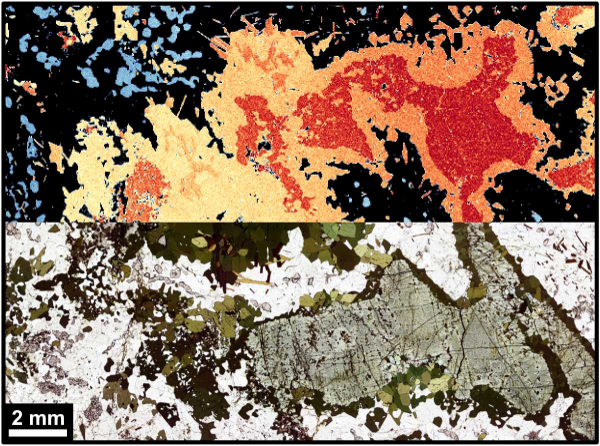
Bottom: optical micrograph of a metagabbro from the Trans-North China Orogen.
Top: quantitative EPMA compositional map of the same area, processed with XMapTools. Diffusion modelling of frozen-in chemical gradients in garnet showed that a reaction long thought to take hundreds of thousands of years formed in days to months. Liu et al. (2025), GCA 394, 106–125. doi:10.1016/j.gca.2025.01.036.
© Lanari, UNIL.
XMapTools: From chemical maps to petrological interpretation
The laboratory is also home to the development of XMapTools, a free, open source and internationally used software package first released in 2014 for processing and interpreting quantitative compositional maps.
XMapTools transforms raw intensity maps acquired by EPMA, SEM, LA-ICP-MS and other analytical techniques into calibrated maps of mineral compositions. Its capabilities include advanced tools for data visualisation, the calculation of mineral structural formulae, mineral modes and local bulk compositions, as well as the integration of compositional maps with advanced phase equilibrium models.
Laboratory users can benefit from direct support throughout the analytical workflow, from data acquisition and map processing to quantitative petrological interpretation and modelling.
Instrument specifications
JEOL JXA-8530F HyperProbe
- Schottky field emission electron gun, providing spatial resolution below 1 µm.
- Five wavelength-dispersive spectrometers (WDS): LIF, PET, TAP, LDE1 and LDE2.
- Height[MR1] large analysing crystals for high analytical sensitivity.
- Elemental analysis from B to U.
- Silicon drift detector (EDS).
- Panchromatic cathodoluminescence detector.
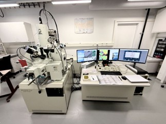
EPMA laboratory, Institute of Earth Sciences (ISTE), University of Lausanne (UNIL).
© Lanari, UNIL.
Sample preparation
Reliable quantitative analysis requires a clean, flat and carefully polished sample surface. Non-conductive materials are generally carbon-coated to ensure electrical conductivity and minimise charging effects during analysis.
Contact

Prof. Pierre Lanari
Head of the lab
pierre.lanari[at]unil.ch
+41 (0)21 692 44 68

Dr. Martin Robyr
Lab Manager
martin.robyr[at]unil.ch
+41 (0)21 692 44 41

